The equine eye includes the eyeball and the surrounding muscles and structures around the eyeball, termed the adnexa.
The eyeball of a horse
The eyeball of the horse is not perfectly spherical, but rather is flattened anterior to posterior. However, research has found the horse does not have a ramped retina, as was once thought.
The wall of the eye is made up of three layers: the internal or nervous tunic, the vascular tunic, and the fibrous tunic.
The nervous tunic (or retina) is made up of cells which are extensions of the brain, coming off the optic nerve.These receptors are light-sensitive, and include cones, which are less light-sensitive, but allow the eye to see color and provide visual acuity, and rod cells, which are more light-sensitive, providing night vision, but only seeing light and dark differences.
Since only two-thirds of the eye can receive light, the receptor cells do not need to cover the entire interior of the eye, and line only the area from pupil to the optic disk.
The part of the retina covered by light-sensitive cells is therefore termed the pars-optica retinae, and the blind part of the eye is termed the pars-ceaca retinae.
The optic disk of the eye, however, does not contain any of these light-sensitive cells, as it is where the optic nerve leaves to the brain, so it is a blind spot within the eye.
The vascular tunic (or uvea) is made up of the choroid, the ciliary body, and the iris. The choroid has a great deal of pigment, and is almost entirely made of blood vessels.
It forms the tapetum lucidum when it crosses over the fundus of the eye, causing the yellowish-green eye shine when light is directed into the animal’s eyes at night.The tapetum lucidum reflects light back onto the retina, allowing for greater absorption in dark conditions.
Eye color in horses
The iris lies between the cornea and the lens, and not only gives the eye its color, but also allows for varying amounts of light to pass through its center hole, which is the pupil
The fibrous tunic consists of the sclera and cornea and protects the eye. The sclera (white of the eye) is made up of elastin and collagen. The cornea (clear covering on the front of the eye) is made up of connective tissue and bathed in lacrimal fluid and aqueous humor, which provides it nutrition, as it does not have access to blood vessels.
The lens of the eye lies posterior to the iris, and is held suspended by the ciliary suspensory ligament and the ciliary muscle, which allows for accommodation of the eye: it allows the lens to change shape to focus on different objects. The lens is made up of onion-like layers of tissue.
 Although usually dark brown, the iris may be a variety of colors, including blue, hazel, amber, and green.
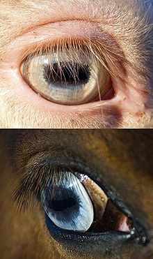
Homozygous cream dilutes (which are double-dilutes) have pale blue eyes (left top), while the blue eyes associated with white markings (left bottom) are a clearer, deeper color.
Blue eyes are not uncommon and are associated with white markings or patterns. The white spotting patterns most often linked to blue eyes are splashed white, frame overo, and sometimes sabino. In the case of horses with white markings, one or both eyes may be blue, or part-blue.
Homozygous cream dilutes, always have light blue eyes to match their pale, cream-colored coats. Heterozygous or single-dilute creams, such as palominos and buckskins, often have light brown eyes. The eyes of horses with the Champagne gene are typically greenish shades: aqua at birth, darkening to hazel with maturity.
As in humans, much of the genetics and etiology behind eye color are not yet fully understood.
Horses have three eyelids
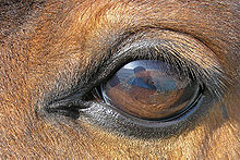 The adnexa of the eye, including the third eyelid (seen in the left corner) is shown in the picture to left. The eyelids are made up of three layers of tissue: a thin layer of skin, which is covered in hair, a layer of muscles which allow the lid to open and close, and the palpebral conjunctiva, which lies against the eyeball. The opening between the two lids forms the palpebral fissue. The upper eyelid is larger and can move more than the lower lid. Unlike humans, horses also have a third eyelid (nictitating membrane) to protect the cornea. It lies on the inside corner of the eye, and closes diagonally over it.
The lacrimal apparatus produces tears, providing nutrition and moisture to the eye, as well as helping to remove any debris that may have entered. The apparatus includes the lacrimal gland and the accessory lacrimal gland, which produce the tears. Blinking spreads the fluid over the eye, before it drains via the nasolacrimal duct, which carries the lacrimal fluid into the nostril of the horse.
The ocular muscles allow the eye to move within the skull.